Overview
The Stem Cell Biology group are particularly interested in understanding the mechanisms of development and maintenance of leukaemia and identifying and validating new therapeutic targets. Another focus of our work is to uncover the mechanisms of normal haematopoietic development to recapitulate this process in vitro for the generation of therapeutic immune cells.
Cytotoxic therapy has been the standard of care over the last 30 years for acute myeloid leukaemias (AMLs). Unfortunately, more often than not, it fails to cure patients, and the five years survival rate is only around 20%. Therefore, there is a pressing need to develop more specific and efficient therapies.
To this end, our laboratory undertakes two integrated research programmes: a) investigating mechanisms driving initiation and maintenance of leukaemia; and b) extending the understanding of normal haematopoietic system development. Our overarching goals are to improve patient outcomes by identifying and validating new therapeutic targets for leukaemia treatment and developing robust protocols for in vitro production of clinical-grade blood cells for adoptive cancer immunotherapies.
We aim to identify and validate new therapeutic targets in acute myeloid leukaemia. We are implementing advanced technologies, including single-cell multi-omics, CRISPR screening, mass spectrometry, bioinformatics, mouse models, organoid cultures and patient-derived samples analyses in these studies. We also aim to improve our understanding of blood cell development in order to better replicate this process in vitro to generate cells for adaptive cellular immunotherapies. Currently, our incomplete understanding of normal in vivo haematopoietic ontogeny limits our capacity to efficiently replicate this process in vitro. The complexity of haematopoietic development has to be comprehensively defined, and the underlying regulatory mechanisms understood to establish effective in vitro protocols that drive differentiation towards specific haematopoietic lineages.
Featured Publications

Single-cell profiling reveals three endothelial-to-hematopoietic transitions with divergent isoform expression landscapes
11th November 2025
Authors unraveling molecular cues driving the generation of specific blood cell types present a comprehensive scRNA-seq dataset that covers three parallel embryonic EHT trajectories.

The small inhibitor WM-1119 effectively targets KAT6A-rearranged AML, but not KMT2A-rearranged AML, despite shared KAT6 genetic dependency
8th October 2024
The epigenetic factors KAT6A and KMT2A interact in normal hematopoiesis to regulate progenitors’ self-renewal. Authors evaluated the potential of different KAT6A therapeutic targeting strategies to alter the growth of KAT6A and KMT2A rearranged AMLs.

Murine AGM single-cell profiling identifies a continuum of hemogenic endothelium differentiation marked by ACE
20th January 2022
Here, the authors construct a comprehensive atlas of the endothelial-to-haematopoietic transition (EHT) continuum, as well as the subaortic niche cells in mouse embryonic aorta using a set of haemogenic endothelium (HE) reporter models.

The Oncogenic Transcription Factor RUNX1/ETO Corrupts Cell Cycle Regulation to Drive Leukemic Transformation
8th October 2018
Leukaemic fusion proteins drive leukaemia by maintaining abnormal transcriptional networks. This study demonstrates the feasibility of epigenomics-instructed screens for identifying oncogene-driven vulnerabilities and their exploitation by repurposed drug approaches.
Developmental Haematopoiesis
Embryonic sites of blood development
In murine embryos, haematopoiesis takes place in several tissues where blood cells are generated and/or undergo maturation. The first haematopoietic progenitors are found extraembryonically at E8.0–E8.5, in the yolk sac blood islands in close proximity to emerging endothelial cells. Once circulation is established, blood cells colonise other developing haematopoietic organs.
Around E10.5, the AGM, placenta, umbilical artery and vitelline artery initiate the generation of blood precursors that, together with yolk sac cells, colonise to the foetal liver rudiment around E11.5. The fetal liver is the major hematopoietic site where blood progenitors expand and/or mature.
Finally, the bone marrow is colonised by precursors from the foetal liver before birth and remains the main haematopoietic niche throughout adult life.

Haematopoietic stem cell generation
HSCs are generated in mice intra-embryonically in the aorta-gonad-mesonephros (AGM) between embryonic day (E) E10.5 to E11.5, where haemogenic endothelium (HE) cells transition to non-adherent haematopoietic cells via a process termed the endothelial-to-haematopoietic transition (EHT).
Specialised HE cells lining the floor of the dorsal aorta (1) undergo major morphologic changes (2) and EHT produces intra-aortic haematopoietic clusters into the lumen of the dorsal aorta and other major arteries (3).
The group made new insights into the mechanisms regulating HSC emergence from the haemogenic endothelium, with implications for the generation of clinically available HSCs.

Meet the group
Here are the current members of the Stem Cell Biology group.
Join our Group
We don’t have any current vacancies here in our group at the moment, but please check out the Institute careers pages here.
Get in touch
Our vision for world leading cancer research in the heart of Manchester
We are a leading cancer research institute within The University of Manchester, spanning the whole spectrum of cancer research – from investigating the molecular and cellular basis of cancer, to translational research and the development of therapeutics.
Our collaborations
Bringing together internationally renowned scientists and clinicians
Scientific Advisory Board
Supported by an international Scientific Advisory Board
Careers that have a lasting impact on cancer research and patient care
We are always on the lookout for talented and motivated people to join us. Whether your background is in biological or chemical sciences, mathematics or finance, computer science or logistics, use the links below to see roles across the Institute in our core facilities, operations teams, research groups, and studentships within our exceptional graduate programme.




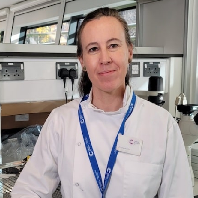
A note from the Group Leader – Georges Lacaud
Our team is composed of members with diverse expertise, fostering a collaborative environment where everyone supports one another and can realise their full potential. We are committed to advancing science while also embracing the excitement and enjoyment that comes with making new discoveries.